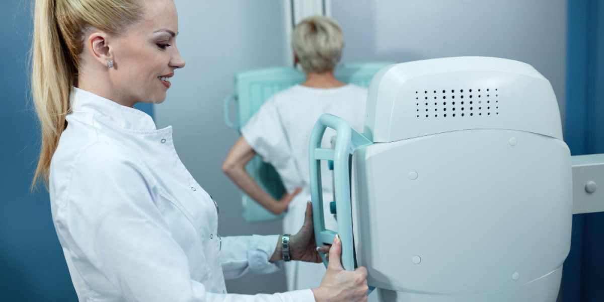
Koning Vera 3D Breast CT Is Changing How Women Get Screened
For a lot of women in the US, the annual mammogram is something they dread more than they look forward to. The compression, the awkward positioning, the anxiety of waiting for results — it adds up. So when a technology comes along that genuinely rethinks how breast imaging works, it's worth paying attention.
The Koning Vera 3D breast CT is doing exactly that. It's not a tweak to existing mammography. It's a fundamentally different approach — one that's already shifting how radiologists and patients think about breast cancer detection.
If you've heard the name but aren't sure what it actually means for you, this is the guide that breaks it down clearly and honestly.
What the Koning Vera System Actually Does
At its core, the Koning Vera is a dedicated breast CT scanner. That means it was designed from the ground up specifically for breast imaging — not adapted from a general-purpose CT machine.
Here's how it works in practice: you lie face-down on a specially designed table with a comfortable opening. The breast hangs naturally into the scanning field, and the system rotates around it to capture hundreds of images from multiple angles. The entire scan takes about 10 seconds per breast.
The result is a true 3D volumetric image of the entire breast — every cubic millimeter of tissue rendered in high resolution, in three dimensions, without distortion or overlap.
Why that 3D difference is significant
Traditional mammography produces 2D images. Even 3D mammography (tomosynthesis) is still a compression-based technique that captures a limited number of angled slices. Koning Vera CT captures the entire breast volume at once, giving radiologists the ability to scroll through every layer of tissue with far greater clarity.
Overlapping tissue — one of the most common reasons tumors get missed in standard mammograms — becomes a non-issue when you're working with a complete 3D dataset. Radiologists can isolate and examine specific regions without the visual noise that flat imaging creates.
The Compression Question: Why It Matters More Than You Might Think
Let's talk about compression, because it's one of the most cited barriers to breast cancer screening compliance in the US.
Studies consistently show that fear of pain and discomfort is a real reason women delay or skip mammograms. For women with dense breast tissue, fibrocystic conditions, or implants, that discomfort can be significant. For women who've had breast surgery or are particularly sensitive, it can be genuinely painful.
No compression breast imaging is not just a comfort upgrade — it has downstream implications for public health. When screening is less intimidating, more women show up. When more women show up, more cancers get caught early. That's not a marketing point; it's a straightforward logical chain.
The Koning Vera Breast CT eliminates compression entirely. The breast is never squeezed, flattened, or held in position. It rests naturally, the scanner does its work, and you're done. That's a meaningful change in the patient experience — particularly for women who've been avoiding regular screening because of how uncomfortable it's been in the past.
Dense Breast Tissue: Where Standard Mammography Falls Short
About 40% of women in the US have dense breast tissue. Dense tissue shows up white on a mammogram. So does cancer. This creates a masking problem where tumors hiding within dense tissue can be extremely difficult to detect — even by experienced radiologists reading high-quality images.
This is one of the most important clinical arguments for a technology like Koning Vera 3D breast CT.
How CT imaging handles density differently
CT imaging assigns different density values to different tissue types. Because the system captures full volumetric data rather than projecting everything onto a flat plane, dense tissue and suspicious masses are far easier to differentiate. Radiologists aren't trying to spot something white against a white background — they're working with a rich, layered dataset that shows tissue characteristics in three dimensions.
For women who've been told they have dense breasts and have been anxious about the reliability of their screenings, this is a genuinely reassuring development. It doesn't mean every imaging center in the US offers it yet, but access is growing.
How It Compares to Other Imaging Options
It's worth being clear-eyed about this: no single imaging modality is perfect for every patient in every situation. Here's how Koning Vera fits into the broader landscape.
Versus traditional mammography
Standard 2D mammography is still the most widely available breast screening tool in the US and has a strong track record. But it has real limitations — particularly for dense tissue and for detecting certain tumor types in their early stages. Koning Vera CT was designed to address precisely those limitations.
Versus MRI
Breast MRI is highly sensitive and is already used for high-risk patients. But it requires IV contrast, takes significantly longer, is considerably more expensive, and is not appropriate for routine annual screening for most women. Dedicated breast CT like the Koning Vera offers many of the three-dimensional advantages of MRI at a fraction of the time and with far less invasiveness.
Versus ultrasound
Ultrasound is useful as a supplemental tool, particularly for evaluating specific areas of concern identified on mammography. It is operator-dependent, doesn't cover the whole breast efficiently, and doesn't provide the comprehensive volumetric data that a breast CT scan delivers.
The breast ct scan powered by the Koning Vera sits in an interesting position — it's more comprehensive than mammography, more accessible than MRI, and more efficient than relying on ultrasound as a supplemental layer.
Radiation: The Question Everyone Has
It's a fair concern and it deserves a straight answer. Any CT imaging involves ionizing radiation. The Koning Vera system is designed to deliver radiation doses comparable to standard mammography — the developers and clinical teams involved in its design were well aware that dose management is a core requirement for a system intended for routine screening use.
That said, radiation exposure in medical imaging is always a conversation to have with your radiologist or physician, who can weigh your individual history, risk factors, and the clinical value of each imaging option. The goal is always the most useful information at the lowest reasonable dose.
Where You Can Access Koning Vera Breast CT in the US
Adoption is growing but still concentrated at specialized imaging centers, academic medical centers, and forward-thinking radiology practices. If you're interested in accessing this technology, the most direct path is talking to your OB-GYN, primary care physician, or a breast imaging specialist and asking specifically whether Koning Vera CT is available in your area or at a referral center.
Women with dense breasts, a family history of breast cancer, prior inconclusive mammograms, or a strong preference for compression-free screening have particularly compelling reasons to explore whether this technology is accessible to them.
Take the Next Step Toward Better Breast Health
You don't have to accept that breast screening has to be uncomfortable or uncertain. Advances like Koning Vera 3D breast CT exist precisely because better is possible — and you deserve access to the best tools available.
Talk to your doctor about whether dedicated breast CT imaging is right for your situation. Ask about facilities in your area that offer it. And if you've been putting off your screening because the experience has been unpleasant in the past, let this be the reason you reconsider. Early detection saves lives, and now there are more comfortable ways to get there.